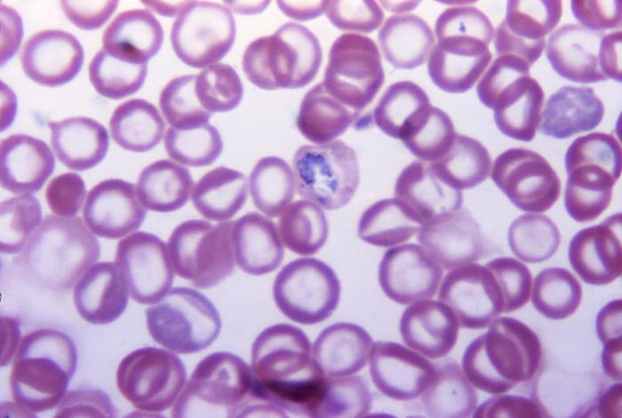
41 Animal Cell Under Light Microscope
Igcse 2014 15 Biology4igcse. In most plant cells the organelles that are visible under a compound light microscope are the cell wall cell membrane cytoplasm central vacuole and nucleus.
Imageanimal cell seen under light microscope.

Animal cell under light microscope. To identify plant and animal cells you must use a microscope with at least 100x magnification power. A brief explanation of the. Animal cells under a light microscope.
Http Www Oncoursesystems Com Images User 9341 10845583 Animal. Plant animal and bacterial cells have smaller components each with a specific function. Every organism composed of one or more cells.
The structures within the cell are referred to as organelles. All cells are categorized in to two groups- Prokaryotic and Eukaryotic. Animal Plant Cells Gcse Science Biology Get To Know Science Youtube Mitochondrion are visible with a light microscope but cant be seen in detail.
Posted 6 years ago. Answer the following questions in your exercise book. Has 5 visible organelles looking through a compound light microscope.
There are one or more cells that form organism. Now up your study game with Learn mode. Cell is a tiny structure and functional unit of a living organism containing various parts known as org.
Structure of Animal Cell and Plant Cell Under Microscope Learn the structure of animal cell and plant cell under light microscope. If you were wondering what is an animal cell below is your answer showing a picture of animal cell. Some of the cell organelles that can be observed under the light microscope include the cell wall cell membrane cytoplasm nucleus vacuole and chloroplasts.
Generalized Structure of an Animal Cell Diagram You know Animal cell structure contains only 11 parts out of the 13 parts you saw in the plant cell diagram because Chloroplast and Cell Wall are available only in a plant cell. Ribosomes are only visible with an electron. Within the cell there is a shape of round with a circular structure of granulated part on the epithelial cells.
Labelled animal cell diagram gcse. There are two categories of cells Eukaryotic and Prokaryotic. Below the basic structure is shown in the same animal cell on the left viewed with the light microscope and on the right with the transmission electron.
Under the microscope animal cells appear different based on the type of the cell. Investigating cells with a light microscope Once slides have been prepared they can be examined under a microscope. Animal cell under the microscope.
Aims of the experiment to use a light microscope to examine animal or plant cells. In this chapter we are making user to control a light microscope remotely using a eukaryotic cell. 14 can you distinguish features of.
You can observe this epithelial animal cell under microscope with high power. Light and electron microscopes allow us to see inside cells. Cell Humans Examples Body.
The nucleus is the control center. A typical animal cell is 1020 μm in diameter which is about one-fifth the size of the smallest particle visible to the naked eye. Meiosis cell division 3d cell cellular division embryo 3d cell animal embryo reproductive health blood cells under microscope the cell cytyoplasm cytoplasm.
You just studied 22 terms. Viewing Animal Cells under a microscope. These cell organelles perform specific functions within the cell.
A cell is the structural and functional unit of life. Real Animal Cell Microscope. Animal cells have a basic structure.
They are all typical elements of a cell. 16 p4 which is photomicrograph of actual animal cells. This shows a generalized animal cell under a light microscope.
3017 animal cell under microscope stock photos vectors and illustrations are available royalty-free. Animal Cell as shown above. The granulated area is the cell Cytoplasm while the huge round part is the Nucleus.
These cell organelles perform specific functions within the cell. It directs all of the cells activities. A cell is the smallest functional and structural entity of life that it is easier observing animal cell under light microscope.
Some of the cell organelles that can be observed under the light microscope include the cell wall cell membrane cytoplasm nucleus vacuole and chloroplasts. Using the labels of Fig. Section 5 Cells View As Single Page.
The structures within the cell are referred to as organelles. Similarly it is asked how do you identify an animal cell. Browse 163 animal cells under microscope stock photos and images available or start a new search to explore more stock photos and images.
In animal cells youll see a round shape with an outer cell membrane and no cell wall. See animal cell under microscope stock video clips. A partial cross section of a worm under a microscope - animal cells under microscope stock pictures royalty-free photos images.
Animal Cell Under Light Microscope Observation. Ziehen die pins an die richtige stelle auf dem bild. The brain of the cell.
However the internal structure and organelles are more or less similar.

